The pituitary gland
Key words: feline, pituitary, adenohypophysis, neurohypophysis
The author apologizes to those with no interest in etymology. They should overlook digressions in that regard.
Abbreviations:
Adrenocorticotropic hormone: ACTH
Corticotropin-like intermediate peptide: CLIP
Pro-opiomelanocortin: POMC
Cerebrospinal fluid: CSF
Thyroid stimulating hormone: TSH
Gonadotrophin releasing hormone: GnRH
Luteinizing hormone LH
Follicle stimulating hormone FSH
Growth hormone releasing hormone: GHRH
Growth hormone inhibiting hormone: GHIH
A slide of a feline pituitary gland; a reminder of normal anatomy and physiology.
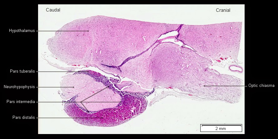
Image size: 891 x 448px.
At right is the optic chiasma, and caudal to it, a ventral portion of the hypothalmus. The third ventricle (a space filled with CSF, dorsal to this part of the hypothalamus) is not visible in this image.
At right is the optic chiasma, and caudal to it, a ventral portion of the hypothalmus. The third ventricle (a space filled with CSF, dorsal to this part of the hypothalamus) is not visible in this image.
Interestingly, the name "pituitary" gland is inappropriate for this structure because the Latin word "pituita" is a term for phlegm; from the ancient belief that this gland produced nasal secretions! Unfortunately, the term hypophysis, less frequently used than pituitary, is less inspiring. It is derived from the Greek words "hypo"(under) and "fusus" (growth) and simply means "undergrowth"; the weeds you find lurking under your shrubs!
During early embryo formation, a pouch-like growth of ectoderm forms in the oral cavity, extending dorsally. It eventually becomes a major part of the pituitary gland.
During early embryo formation, a pouch-like growth of ectoderm forms in the oral cavity, extending dorsally. It eventually becomes a major part of the pituitary gland.
| It may be surprising to read that this up-growth is from the ectoderm, not the endoderm because the gut is mostly of endodermal origon. However, the part of the gut that forms this up-growth is from a small section of the ectoderm that invaginates into the area of the oral cavity. This is similar to the genitals, where the cranial portion of the vagina is endodermal in origin and the vestibule, of ectodermal origin. |
This up-growth of tissue was named for the German physician and embryologist who first described it during the late Victorian era. Many readers will recognize his name: Martin Heinrich Rathke.
Rathke's pouch grows dorsally and is met by an invagination of tissue growing ventrally from the neural ectoderm, just caudal to Rathke's pouch. These two structures embrace and fuse, with the dorsal part of Rathke's pouch surrounding the tube-like neck of the neural growth, hence its name: the pars tuberalis. As this process of carnal coupling continues, the distal part of Rathke's pouch (distal to the brain i.e. the pars distalis) detaches itself from the roof of the oral cavity and the early pituitary gland is formed.
| Interestingly, it appears as though Rathke first described the development of this pouch in snakes, not mammals. This is evident from a translation of one his early publications on the subject: M. H. Rathke. Entwicklungsgeschichte der Natter (Coluber natrix). Königsberg, Bornträger, 1839. (Evolution of the snake (a nasty poisonous viper found in the Southern US). |
Rathke's pouch develops into the anterior (or glandular) part of pituitary. As a result, it is also known as the adenohypophysis, "aden" being the Greek word for gland. In contrast, the posterior pituitary manufactures no secretions (its products are formed in the hypothalamus); it only stores them. For that reason, the posterior pituitary lacks the moniker "aden". Also it is entirely of neural origin, so it is simply known as the neurohypophysis.
| As any veterinary student will know, the pituitary gland is encompassed in a bony cavity at the base of the skull. This was named the sella turcica in the inventive mind of some anatomist because of its resemblance to a Turkish saddle from Latin words sella (saddle) and turcica (Turkish) probably because of an obsession with all things Turkish at this time, just before the decline of thee Ottoman empire. This is an old bit of veterinary trivia but digressing further, readers may be surprised to know that the sella turcica was also known as the "ephippion" at about this time; ephippion being the Latin term for a horse blanket! This comes from examining Samuel George Morton's book on human anatomy published in 1849: An Illustrated System of Human Anatomy, Special, General and Microscopic. So, horse blanket or Turkish saddle, the shape of the structure obviously struck similar chords in those who first examined it. One final digression: In fetuses where the sella turcica is deformed or absent due to heritable conditions or the ingestion of toxic plants (Veratrum californicum), the anterior pituitary is damaged accordingly. In domestic ruminants, this is common & leads to prolonged gestation because of a deficiency of ACTH (see below). |
It is fortunate that a local portal system has evolved between the hypothalamus and the pituitary gland, delivering hypothalamic releasing hormones directly to the pituitary gland. Were it not for this, releasing hormones would enter the systemic circulation and become diluted then partially catabolized by the liver. Eventually they would reach the pituitary gland in extremely low concentrations and a good deal later than otherwise. Fortunately this system evolved before we failed to reproduce and became extinct.
|
In the images below, are the cells in the adenohypophysis that respond to these releasing hormones. Also shown are segments of the neurohypopysis and pars intermedia. Although the pars intermedia also arises from Rathke's pouch, it only develops into a thin band of tissue, almost isolated from the rest of the pars distalis. As one can see below, it also has a distinct appearance, with a striking predominance of basophils (producing POMC) and an a complete absence of acidophils.

In the adenohypophysis, the acidic pH of glycoproteins in the cytoplasm of some cell attracts basic stains. Therefore these cells are known as basophils. In this image, hematoxylin, a common bluish-purple basic stain, identifies cells that contain glycoproteins. These glycoproteins include LH, FSH, TSH and POMC. It should be noted that POMC is indeed a glycoprotein although ACTH (one of its principal breakdown products) is a polypeptide hormone.
| POMC is the main product of the pars intermedia too, so it is not surprising to see that it takes on an exclusive basophilic appearance. The pars intermedia is poorly developed in some species (primates and humans) but well developed in domestic species. In horses, the pars intermedia and POMC take on special significance when adenomas form there, producing a Cushings-like syndrome known as pars pituitary intermedia sysfunction or PPID. |
Close examination of the pars distalis reveals the following:

Image size: 1364 x 778px
Note the predominance of acidophils, staining with eosin, an acidic stain attracted to the basic polypeptide hormones in these cells. The vast majority of these acidophils produce GH. The others produce prolactin.
There is growing evidence that some cells in the adenohypophysis may produce several hormones and may not be restricted to the production of a single hormone as is classically taught. For example, a basophil producing TSH may also produce LH and even prolactin, a hormone usually found in acidophils. Also, it is commonly stated that chromophobes produce no hormones yet they must have essential functions. Clearly therefore, there is still a great deal to discover about the hypothalamic pituitary axis.
One final note: Although many years have passed since the discovery of axonal delivery of oxytocin and vasopressin (ADH) from the hypothalamus to the neurohypophysis, these two hormones still appear to be the only hormones secreted by that part of the pituitary gland.
Selected references/sources:
http://emedicine.medscape.com/article/1899167-overview#aw2aab6b3
Van Poppel, H. and Klotz, L. 2012 Gonadotropin-releasing hormone: An update review of the antagonists
versus agonists. Int. J. Urology. 19:594-601
Kiarisemail, H. et al. 2011 Growth hormone-releasing hormone: not only a neurohormone. Trends Endocrinol. and Metab. 22: 311–317
http://flipper.diff.org/app/items/info/1730
Roseweir, A.K. and Millar, R.P. The role of kisspeptin in the control of gonadotrophin secretion.
Human Reprod.Update 15:203-212.